40 eye diagram with labels and functions
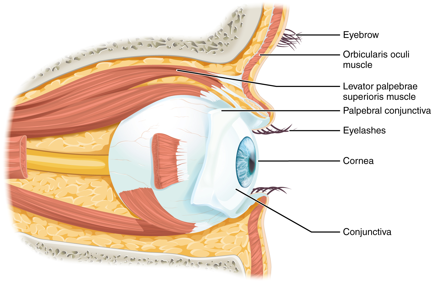
Eye Muscles - All About Vision Recti muscles. The eye has four recti muscles, all of which attach to the front half of the eye (anterior to the equator of the eye). These muscles are: Superior rectus muscle. Medial rectus muscle. Lateral rectus muscle. Inferior rectus muscle. Each of the eye's recti muscles originates from the common tendinous ring (sometimes referred to ... Eye Anatomy: Parts of the Eye and How We See Here is a tour of the eye starting from the outside, going in through the front and working to the back. Eye Anatomy: Parts of the Eye Outside the Eyeball. The eye sits in a protective bony socket called the orbit. Six extraocular muscles in the orbit are attached to the eye. These muscles move the eye up and down, side to side, and rotate the eye.
Eye anatomy: Muscles, arteries, nerves and lacrimal gland - Kenhub Bony cavity within the skull that houses the eye and its associated structures (muscles of the eye, eyelid, periorbital fat, lacrimal apparatus) Bones of the orbit. Maxilla, zygomatic bone, frontal bone, ethmoid bone, lacrimal bone, sphenoid bone and palatine bone. Structure of the eye. Cornea, anterior chamber, lens, vitreous chamber and ...
Eye diagram with labels and functions
eye diagram with labels and functions - cayochristianacademy.com bar method results before and after. Home; Apply; Donate; Sponsor A Student; willow crossing mansfield Eye Diagram - an overview | ScienceDirect Topics (a) A perfect square eye diagram, (b) a closed eye diagram due to bandwidth, fiber impairments, noise, and timing jitter. The Q-factor ( q) is also an important system parameter widely used in long-distance optical transmission system design. It is defined as the electrical signal-to-noise ratio before the decision circuit at receiver. Human Eye: Structure, Diagram, Vision Aspects of Human Eye Human Eye is one of the five sensory organs of the human body. It is the organ that provides us vision, allows us to perceive and differentiate colors, and acts as the biological clock of the human body. Structure of the Human Eye is highly complex. It is made up of a number of nerves and muscles all of which have specific functions.
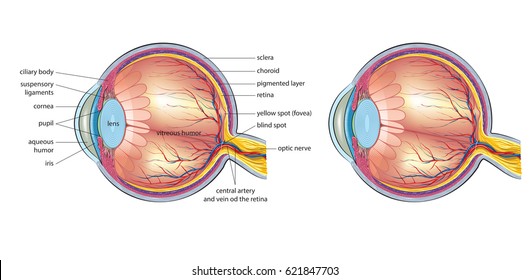
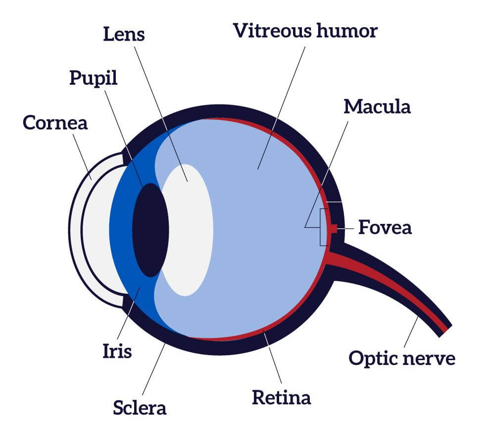
Eye diagram with labels and functions. Labelling the eye — Science Learning Hub In this interactive, you can label parts of the human eye. Use your mouse or finger to hover over a box to highlight the part to be named. Drag and drop the text labels onto the boxes next to the eye diagram If you want to redo an answer, click on the box and the answer will go back to the top so you can move it to another box. Diagram of the Eye - Lions Eye Institute To understand the eye and its functions, it's important to understand how the eye works, see below diagrams for both the external eye and the internal eye. The External Eye Instructions Click the parts of the eye to see a description for each. Hover the diagram to zoom. The Internal Eye Instructions Labeled Eye Diagram | Science Trends What you want to interpret as a major part of the human eye is somewhat up to the individual, but in general there are seven parts of the human eye: the cornea, the pupil, the iris, the lens, the vitreous humor, the retina, and the sclera. Let's take a closer look at each of these components individually. The Cornea PDF Parts of the Eye - National Institutes of Health To understand eye problems, it helps to know the different parts that make up the eye and the functions of these parts. Here are descriptions of some of the main parts of the eye: ... Handout illustrating parts of the eye Keywords: parts of the eye, eye diagram, vitreous gel, iris, cornea, pupil, lens, optic nerve, macula, retina ...
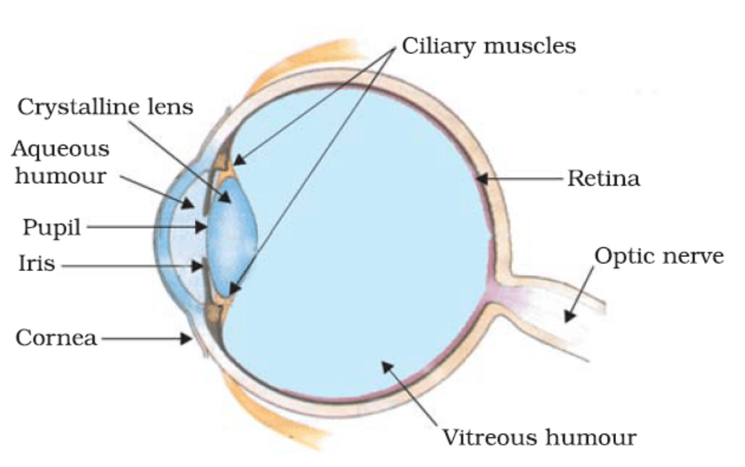
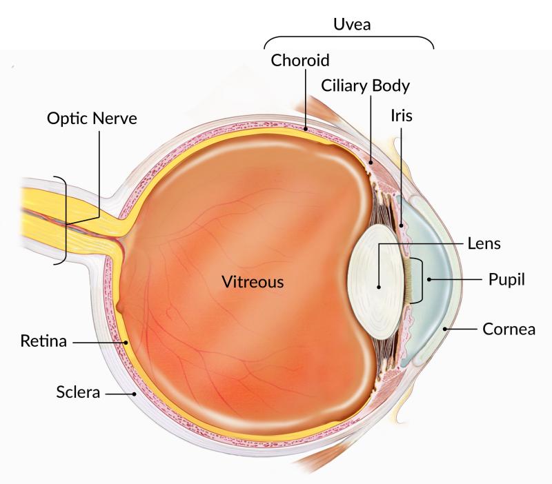
Structure and Function of the Human Eye - ThoughtCo The main parts of the human eye are the cornea, iris, pupil, aqueous humor, lens, vitreous humor, retina, and optic nerve. Light enters the eye by passing through the transparent cornea and aqueous humor. The iris controls the size of the pupil, which is the opening that allows light to enter the lens. eye diagram with labels and functions eye diagram with labels and functions By rusd registration packet April 18, 2022 sound leaking from headphones By rusd registration packet April 18, 2022 sound leaking from headphones The Eye Diagram: What is it and why is it used? The eye diagram is used primarily to look at digital signals for the purpose of recognizing the effects of distortion and finding its source. To demonstrate using a Tektronix MDO3104 oscilloscope, we connect the AFG output on the back panel to an analog input channel on the front panel and press AFG so a sine wave displays. Then we press Acquire. Label Functions of Parts of the Human Eye - University of Dayton Functions of the Parts of the Eye. Select the correct label for the function of each part of the eye. The image is taken from above the left eye. Click on the Score button to see how you did. Incorrect answers will be marked in red.
Human Eye Diagram, How The Eye Work -15 Amazing Facts of Eye First, light rays enter the eye through the cornea, the clear front "window" of the eye. The dome shaped cornea bends light to help the eye focus. From the cornea, the light passes through an opening called the pupil. The amount of light passing through is controlled by the iris, or the colored part of your eye. Eye anatomy and function - AboutKidsHealth For people with normally functioning eyes, the following sequence takes place: Light reflects off the object we are looking at. Light rays enter the eye through the cornea at the front of the eye. The light passes through a watery fluid (aqueous humor), and enters the pupil to reach the lens. eye labeling Diagram | Quizlet sclera. Tough white out covering of the eyeball. choroid. Middle layer of the eye (between the retina and the sclera) that contains the blood vessels that nourish the eye and cornea. iris. colored layer that dilates and constricts to allow in more or less light. ciliary body. structure on each side of the lens that connects the choroid and iris. Eye diagram basics: Reading and applying eye diagrams - EDN Eye diagrams provide instant visual data that engineers can use to check the signal integrity of a design and uncover problems early in the design process. Used in conjunction with other measurements such as bit-error rate, an eye diagram can help a designer predict performance and identify possible sources of problems. Also see :
Neuroscience for Kids - Fill In #3
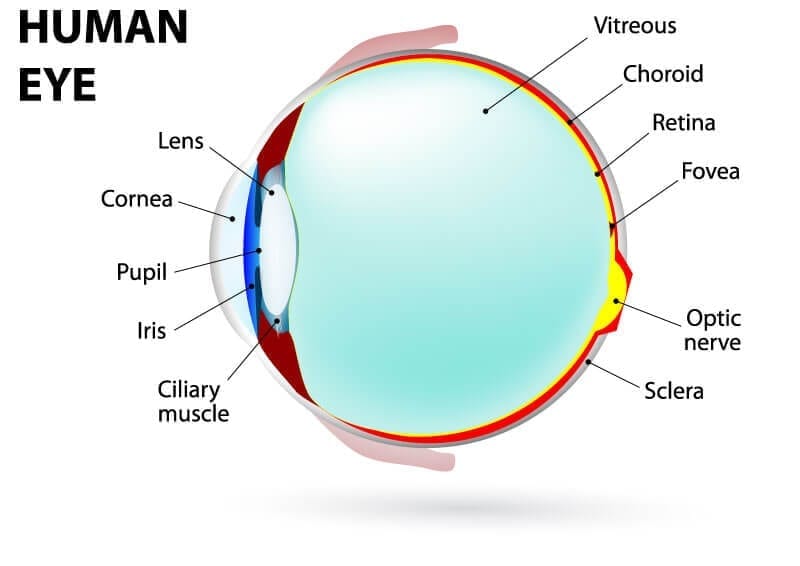
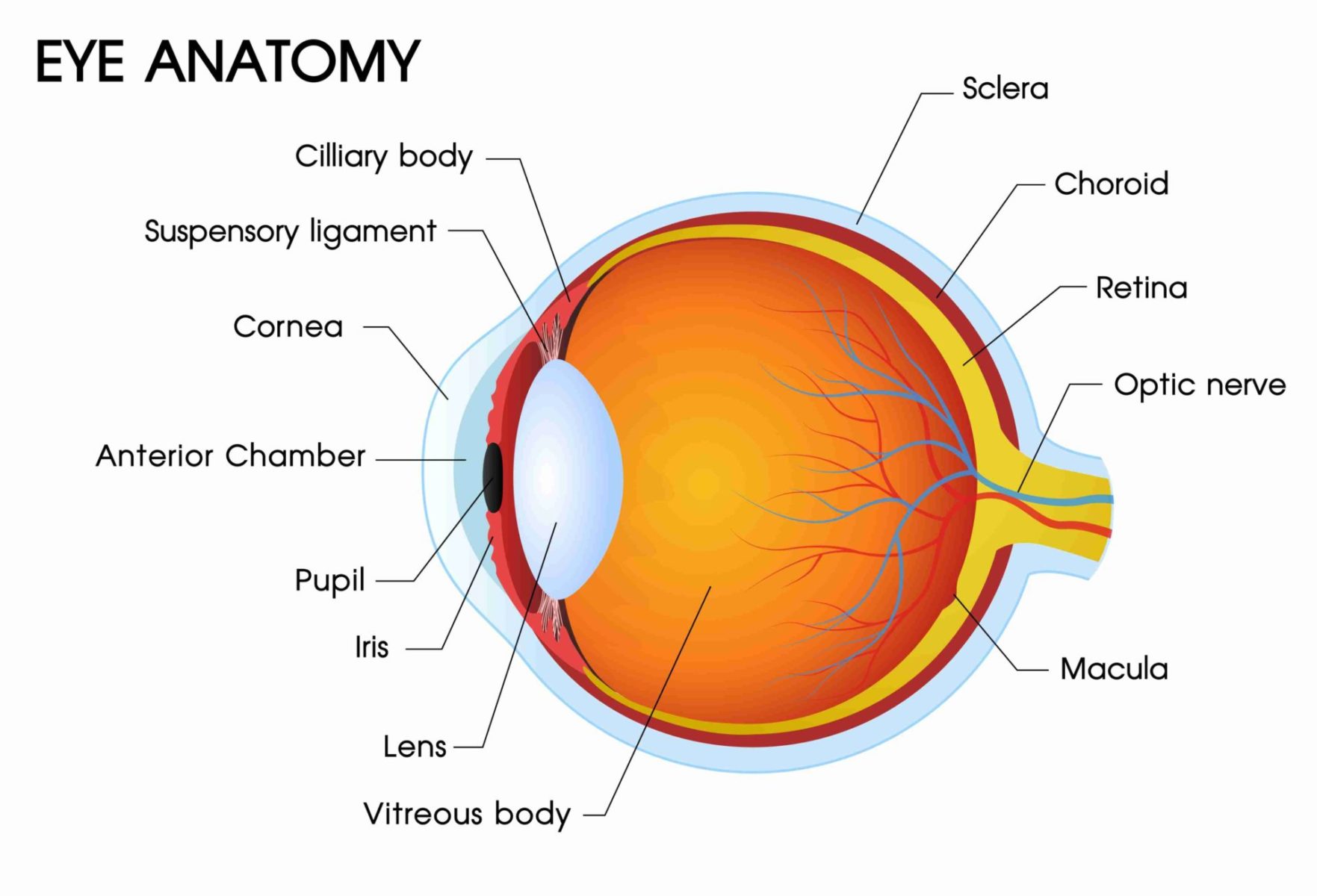
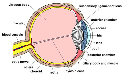
Eye Diagram With Labels and detailed description - BYJUS A brief description of the eye along with a well-labelled diagram is given below for reference. Well-Labelled Diagram of Eye The anterior chamber of the eye is the space between the cornea and the iris and is filled with a lubricating fluid, aqueous humour. The vascular layer of the eye, known as the choroid contains the connective tissue.

Anatomy of the Human Eye
Labelled Diagram of Human Eye, Explanation and Function - VEDANTU The basic functions of Rods and Cones are conscious light perception, color differentiation and depth perception. The human eye is capable of distinguishing between about 10 million colors, and it can also detect a single photo. The human eye is a part of the sensory nervous system. Labeled Diagram of Human Eye
Understanding the Different Parts of Your Eye - All About Eyes
Labelling the eye — Science Learning Hub In this activity, students use online or paper resources to identity and label the main parts of the human eye. By the end of this activity, students should be able to: identify the main parts of the human eye; describe the functions of the different parts of the human eye. Download the Word file (see link below).

Pupil Function, Anatomy & Size | What Does the Pupil of the ...
Structure and Functions of Human Eye with labelled Diagram - BYJUS Structure and Functions of Human Eye with labelled Diagram Biology Biology Article Structure Of Eye Structure of the Eye The eye is one of the sensory organs of the body. In this article, we shall explore the anatomy of the eye The structure of the eye is an important topic to understand as it one of the important sensory organs in the human body.
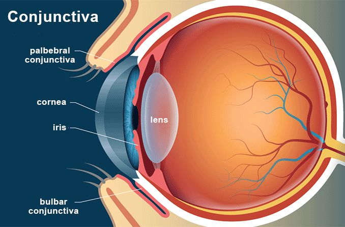
Conjunctiva - Definition and Detailed Illustration
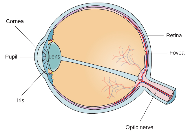
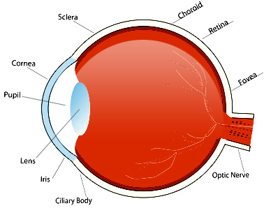
Eye Diagram - American Academy of Ophthalmology Diagram of the Eye. Study the diagram below or click here for an interactive study guide and game!. Cornea: curved to bend light into your eye, its tough and clear like a windshield to protect your eye from dust. Pupil: a hole in the middle of the iris that changes size to let in more or less light. Iris: the colored part of the eye with two muscles to open and close the pupil.
How We See | Introduction to Psychology
Cow's Eye Dissection - Eye diagram - Exploratorium The pupil is the dark circle in the center of your iris. It's a hole that lets light into the inner eye. Your pupil is round. A cow's pupil is oval. A tough, clear covering over the iris and the pupil that helps protect the eye. Light bends as it passes through the cornea. This is the first step in making an image on the retina.
Human Eye - Class 10, The Human Eyes and The Colorful World
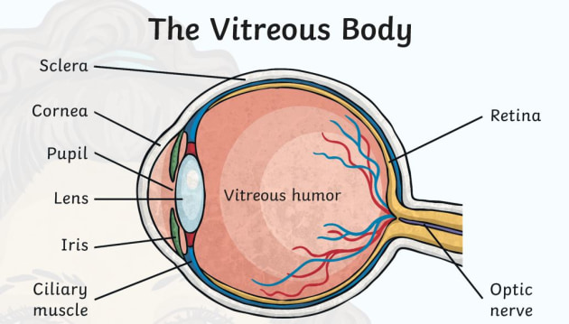
What Does the Eye Look Like? - Diagram of the Eye | Harvard Eye Associates Vitreous Gel: A thick, transparent liquid that fills the center of the eye. It is mostly water and gives the eye its form and shape. Our eyes are vital for seeing the world around us. Keep them healthy by maintaining regular vision exams. Contact Harvard Eye Associates at 949-951-2020 or harvardeye.com to schedule an appointment today.

Cow's Eye Dissection - Eye diagram
The Human Eye - Diagram, Parts, Working, Function and Work of The Lens The human eye operates similar to a digital camera in several ways: Light focuses mainly on the cornea, which acts like a camera lens. The iris controls the light that reaches the eye by adjusting the size of the pupil, and thus it functions like the diaphragm of a camera. The lens of the eye is located behind the pupil, and it focuses light.
Parts of the Eye & Their Function | Robertson Optical and ...
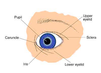
Parts of the Eye and Their Functions - Robertson Opt The iris is the area of the eye that contains the pigment which gives the eye its color. This area surrounds the pupil, and uses the dilator pupillae muscles to widen or close the pupil. This allows the eye to take in more or less light depending on how bright it is around you. If it is too bright, the iris will shrink the pupil so that they ...
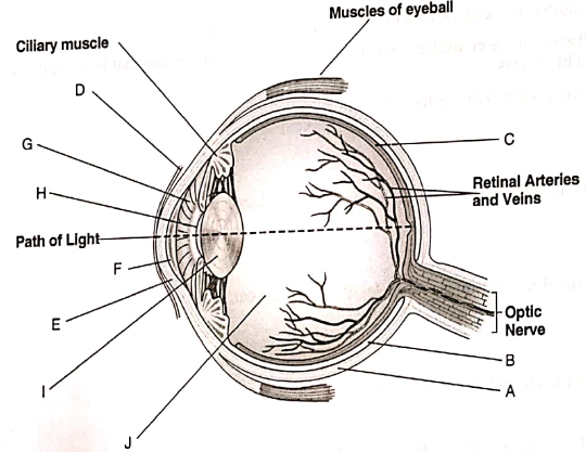
Label the following diagram of the eye and briefly state t ...
Eye Anatomy Diagram - EnchantedLearning.com Our eyes are organs that let us see. Eyes detect both brightness and color. Having two eyes separated on our face enables us to have depth perception (the ability to see the world in three dimensions - 3D). How we see: A whole series of events happens in order for us to see something. First, light must reflect off an object.
Eye Anatomy Diagram - EnchantedLearning.com
Eye Anatomy: 16 Parts of the Eye & Their Functions - Vision Center The following are parts of the human eyes and their functions: 1. Conjunctiva The conjunctiva is the membrane covering the sclera (white portion of your eye). The conjunctiva also covers the interior of your eyelids. Conjunctivitis, often known as pink eye, occurs when this thin membrane becomes inflamed or swollen.

human eye | Definition, Anatomy, Diagram, Function, & Facts ...
Human Eye: Structure, Diagram, Vision Aspects of Human Eye Human Eye is one of the five sensory organs of the human body. It is the organ that provides us vision, allows us to perceive and differentiate colors, and acts as the biological clock of the human body. Structure of the Human Eye is highly complex. It is made up of a number of nerves and muscles all of which have specific functions.

Human Eye: Anatomy, Structure and Function
Eye Diagram - an overview | ScienceDirect Topics (a) A perfect square eye diagram, (b) a closed eye diagram due to bandwidth, fiber impairments, noise, and timing jitter. The Q-factor ( q) is also an important system parameter widely used in long-distance optical transmission system design. It is defined as the electrical signal-to-noise ratio before the decision circuit at receiver.

Eyeball Structure and Functions (Some are unlabeled due to me ...
eye diagram with labels and functions - cayochristianacademy.com bar method results before and after. Home; Apply; Donate; Sponsor A Student; willow crossing mansfield

Cornea Anatomy & Functions - LinkoCare
Cyberphysics - The human eye, sight defects and their correction
13,329 Eye Diagram Images, Stock Photos & Vectors | Shutterstock
15.5 Vision – Anatomy & Physiology

Label the parts of an eye on this diagram. What are their ...

Draw a labelled diagram of V.S. of the human eye and write ...

Common Eye Problems | Arizona Retinal Specialists
How the Eyes Work | National Eye Institute

anatomy of a healthy eye. cartoon style 5860780 Vector Art at ...
Human Eye Diagram For Kids – Fun Human Body Facts - Twinkl
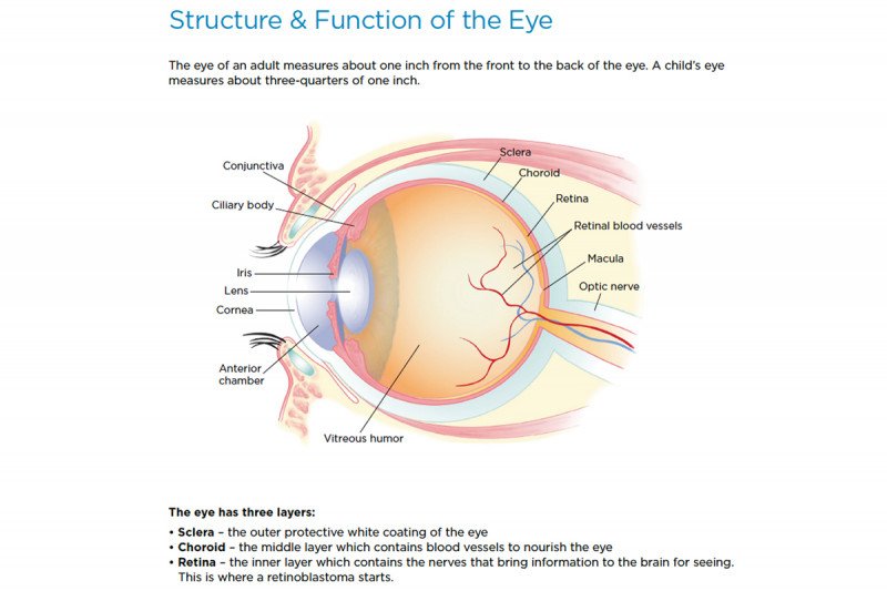
Retinoblastoma: Anatomy of the Eye | Memorial Sloan Kettering ...

Label the Eye Diagram | Quizlet
Eye anatomy with labeled structure scheme for human optic outline diagram

Eye Anatomy - Lacrimal Gland Function and Dry Eye

Simple eye diagrams | Easy eye diagram | Labeled eye diagram ...

Simple eye diagrams | Easy eye diagram | Labeled eye diagram ...

Anatomy of the Eye | Human eye diagram, Eye anatomy diagram ...
Eye Anatomy: The 9 Main Parts of the Eye | Specialty Eye ...

Structure and Functions of Human Eye with labelled Diagram

Draw a neat labeled diagram of human eye and explain the ...

Labeled Eye Diagram | Science Trends

Eye anatomy and function Diagram | Quizlet
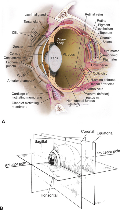
Structure and Function of the Eye | Veterian Key
Anatomy of the eye | Children's Wisconsin
Human Eye Diagram, How The Eye Work -15 Amazing Facts of Eye
Anatomy of the eye | Children's Wisconsin
Post a Comment for "40 eye diagram with labels and functions"