39 compound microscope diagram without labels
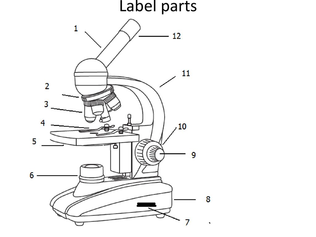
Compound Microscope: Parts of Compound Microscope - BYJUS (A) Mechanical Parts of a Compound Microscope 1. Foot or base It is a U-shaped structure and supports the entire weight of the compound microscope. 2. Pillar It is a vertical projection. This stands by resting on the base and supports the stage. 3. Arm The entire microscope is handled by a strong and curved structure known as the arm. 4. Stage Electron microscope - Wikipedia An electron microscope is a microscope that uses a beam of accelerated electrons as a source of illumination. As the wavelength of an electron can be up to 100,000 times shorter than that of visible light photons, electron microscopes have a higher resolving power than light microscopes and can reveal the structure of smaller objects.. Electron microscopes use shaped magnetic …
Looking at the Structure of Cells in the Microscope A typical animal cell is 10–20 μm in diameter, which is about one-fifth the size of the smallest particle visible to the naked eye. It was not until good light microscopes became available in the early part of the nineteenth century that all plant and animal tissues were discovered to be aggregates of individual cells. This discovery, proposed as the cell doctrine by Schleiden and …

Compound microscope diagram without labels
Label the microscope — Science Learning Hub Use this interactive to identify and label the main parts of a microscope. Drag and drop the text labels onto the microscope diagram. eye piece lens diaphragm or iris coarse focus adjustment stage base fine focus adjustment light source high-power objective Download Exercise Tweet Solved Label the image of a compound light microscope using - Chegg Expert Answer. 100% (17 ratings) Transcribed image text: Label the image of a compound light microscope using the terms provided. Compound Microscope Parts - Labeled Diagram and their Functions Labeled diagram of a compound microscope Major structural parts of a compound microscope There are three major structural parts of a compound microscope. The head includes the upper part of the microscope, which houses the most critical optical components, and the eyepiece tube of the microscope.
Compound microscope diagram without labels. Compound Microscope: Basics, Functionality, and Uses The word compound means multiple, mix, or a combination of both. A compound microscope is a microscope which uses more than one lens. Devised with a system of combination of lenses, a compound microscope consists of two optical parts, namely the objective lens and the ocular lens. The objective lens uses short focal length to enlarge an image. Biopython - Quick Guide - tutorialspoint.com Create a Track for each track you want on the diagram, and add GraphSets and FeatureSets to the tracks you require. Create a Diagram, and add the Tracks to it. Tell the Diagram to draw the image. Write the image to a file. Let us take an example of input GenBank file − What is a Compound Microscope? - New York Microscope Company A compound microscope is an instrument that is used to view magnified images of small specimens on a glass slide. It can achieve higher levels of magnification than stereo or other low power microscopes and reduce chromatic aberration. It achieves this through the use of two or more lenses in the objective and the eyepiece. Compound Microscope- Definition, Labeled Diagram, Principle, Parts, Uses Alternatively, the magnification of the compound microscope is given by: m = D/ fo * L/fe where, D = Least distance of distinct vision (25 cm) L = Length of the microscope tube fo = Focal length of the objective lens fe = Focal length of the eye-piece lens Parts of a Compound Microscope Eyepiece And Body Tube.
A Study of the Microscope and its Functions With a Labeled Diagram ... Compound Microscope Diagram The compound microscope uses light for illumination. Some compound microscopes make use of natural light, whereas others have an illuminator attached to the base. The specimen is placed on the stage and observed through different lenses of the microscope, which have varying magnification powers. Parts of a microscope with functions and labeled diagram - Microbe Notes Figure: Diagram of parts of a microscope There are three structural parts of the microscope i.e. head, base, and arm. Head - This is also known as the body. It carries the optical parts in the upper part of the microscope. Base - It acts as microscopes support. It also carries microscopic illuminators. Labelled Diagram of Compound Microscope The below mentioned article provides a labelled diagram of compound microscope. Part # 1. The Stand: The stand is made up of a heavy foot which carries a curved inclinable limb or arm bearing the body tube. The foot is generally horse shoe-shaped structure (Fig. 2) which rests on table top or any other surface on which the microscope in kept. 16 Parts of a Compound Microscope: Diagrams and Video The 16 core parts of a compound microscope are: Head (Body) Arm Base Eyepiece Eyepiece tube Objective lenses Revolving Nosepiece (Turret) Rack stop Coarse adjustment knobs Fine adjustment knobs Stage Stage clips Aperture Illuminator Condenser Diaphragm Video: Parts of a compound Microscope with Diagram Explained
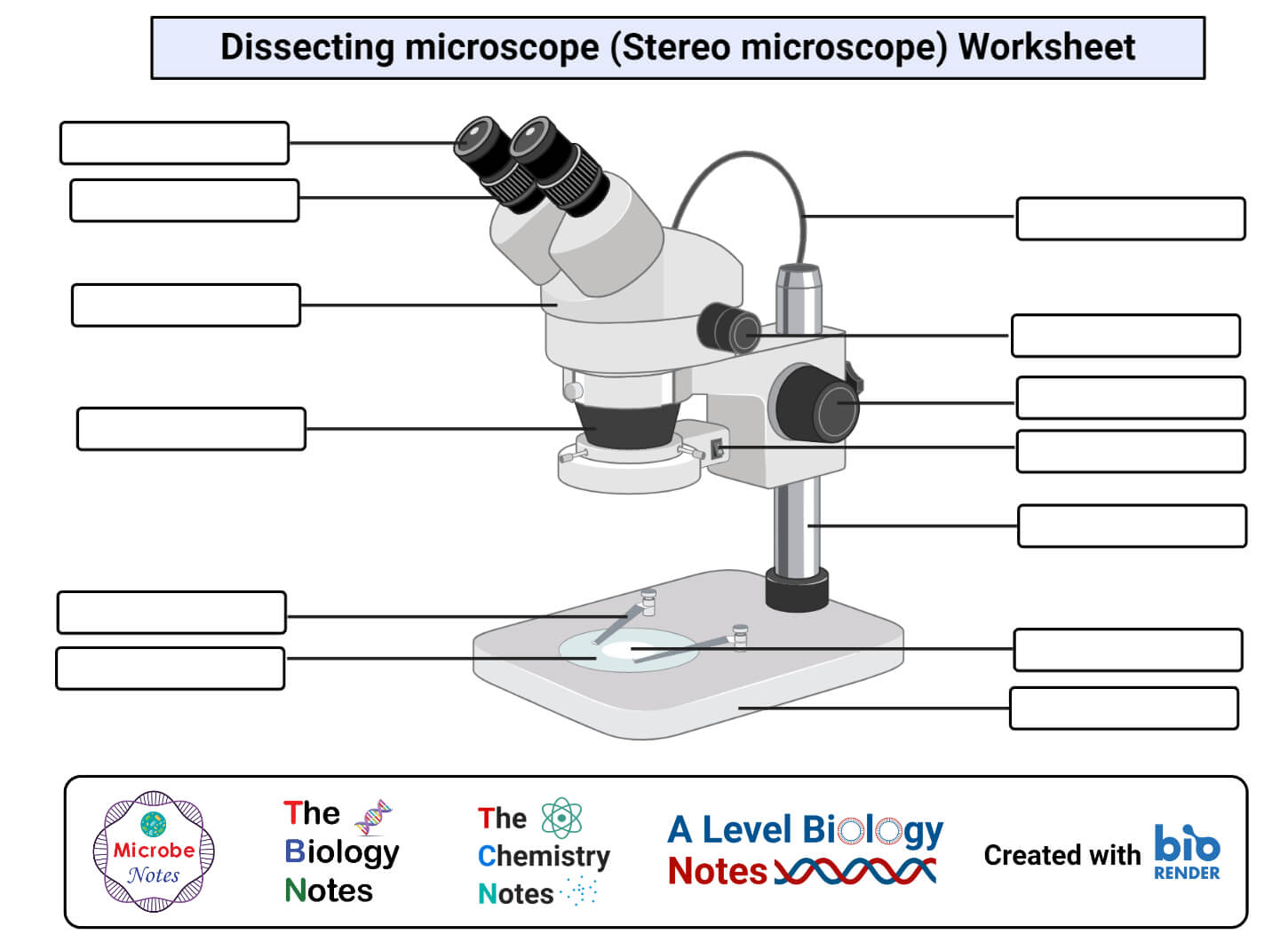
Labeling the Parts of the Microscope | Microscope World Resources Labeling the Parts of the Microscope This activity has been designed for use in homes and schools. Each microscope layout (both blank and the version with answers) are available as PDF downloads. You can view a more in-depth review of each part of the microscope here. Download the Label the Parts of the Microscope PDF printable version here. Parts of Stereo Microscope (Dissecting microscope) – labeled diagram ... If you would like to learn optical components of a compound microscope, please visit Compound Microscope Parts – Labeled Diagram and their Functions, and this article. How to use a stereo (dissecting) microscope. Follow these steps to put your stereo microscopes in work: 1. Set your microscope on a tabletop or other flat sturdy surface where ... How to Use a Compound Microscope: 11 Steps (with Pictures) - wikiHow Focus the microscope. Looking through the eyepiece, arrange the illuminator and the diaphragm to reach the most comfortable level of light. Move the specimen slide so that the image is in the center of your view. [10] Arrange the illuminator until you've arrived at a comfortable level of light. Working Principle and Parts of a Compound Microscope (with Diagrams) It holds the stage, body tube, fine adjustment and coarse adjustment. 5. Body Tube: It is usually a vertical tube holding the eyepiece at the top and the revolving nosepiece with the objectives at the bottom. The length of the draw tube is called 'mechanical tube length' and is usually 140-180 mm (mostly 160 mm). 6.
Dissecting Stereo Microscope Parts and Functions
PDF AN INTRODUCTION TO THE COMPOUND MICROSCOPE - Rowan University microscope in an upright position using both hands. **When carrying the microscope, place one hand on the base and the other hand around the arm. **DO NOT PLACE THE MICROSCOPE IN AN UPSIDE DOWN POSITION. PIECES WILL FALL OUT. **Keep microscope away from the edge of the bench, particularly when not in use. **Make sure power cords are out of the way.

Microscope Labeling
Fluorescence - Wikipedia The chemical compound responsible for this fluorescence is matlaline, which is the oxidation product of one of the flavonoids found in this wood. [1] In 1819, Edward D. Clarke [5] and in 1822 René Just Haüy [6] described fluorescence in fluorites , Sir David Brewster described the phenomenon for chlorophyll in 1833 [7] and Sir John Herschel ...

Compound Microscope Parts, Functions, and Labeled Diagram ...
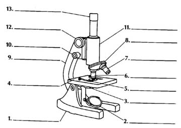
Diagram of a Compound Microscope - Biology Discussion Diagram of a Compound Microscope Article Shared by ADVERTISEMENTS: In this article we will discuss about:- 1. Essential Parts of Compound Microscope 2. Magnification of the Image of the Object by Compound Microscope 3. Resolution Power 4. Method for Studying Microbes 5. Measurement of the Size of Objects. Essential Parts of Compound Microscope:

Compound Microscope: Parts of Compound Microscope
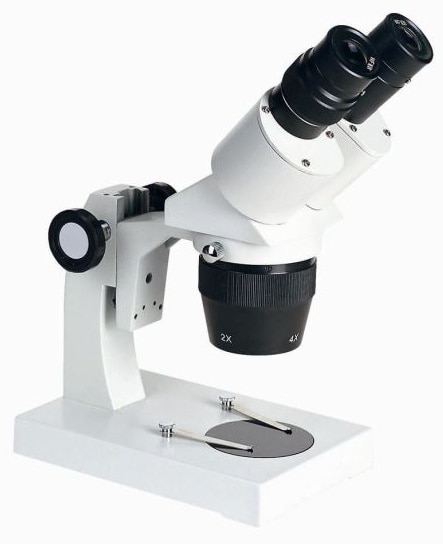
Simple Microscope - Parts, Functions, Diagram and Labelling Picture 1: The image above is a stereo microscope. Image source: made-in-china.com Picture 2: The image above is a confocal microscope. Image source:thorlabs.com Picture 3: The image above is parts of scanning electron microscope. Image source:britannica.com Picture 4: The picture is a transmission electron microscope. Image source: ysjournal.com
Multiple Choice Quiz on Compound Microscope Parts and Functions
Compound Microscope - Types, Parts, Diagram, Functions and Uses Compound microscope - It is an optical instrument consists of two convex lenses of short focal lengths primarily used for observing a highly magnified image of minute objects. Lenses Simple microscope - It has a convex lens. It uses only one lens to magnify objects. An example of a simple microscope is a magnifying glass.

Parts of a Compound Microscope - Labeled (with diagrams ...
Label Microscope Diagram - EnchantedLearning.com Using the terms listed below, label the microscope diagram. arm - this attaches the eyepiece and body tube to the base. base - this supports the microscope. body tube - the tube that supports the eyepiece. coarse focus adjustment - a knob that makes large adjustments to the focus. diaphragm - an adjustable opening under the stage, allowing ...
Compound Microscope Parts, Functions, and Labeled Diagram ...
Parts of a Compound Microscope (And their Functions) - Scope Detective List of Microscope Parts and their Functions. 1. Ocular Tubes (Monocular, Binocular & Trinocular) The ocular tubes, are to tubes that lead from the head of the microscope out to your eyes. On the end of the ocular tubes are usually interchangeable eyepieces (commonly 10X and 20X) that increase magnification.

Parts of the Microscope with Labeling (also Free Printouts ...
On-chip beam rotators, adiabatic mode converters, and … 7.7.2022 · Low-loss-integrated optical waveguides have indispensable roles in a wide range of significant modern technologies, such as integrated photonic chips 1,2,3, quantum applications 4,5,6,7,8,9, and ...

Label the microscope — Science Learning Hub
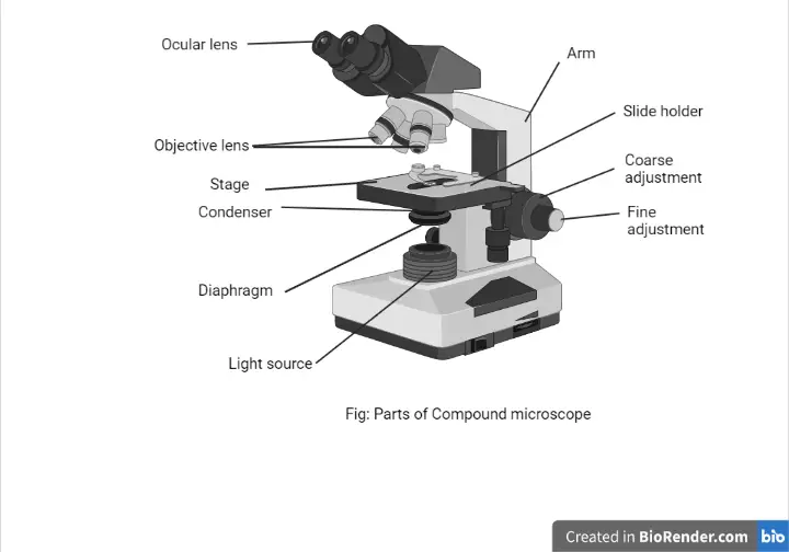
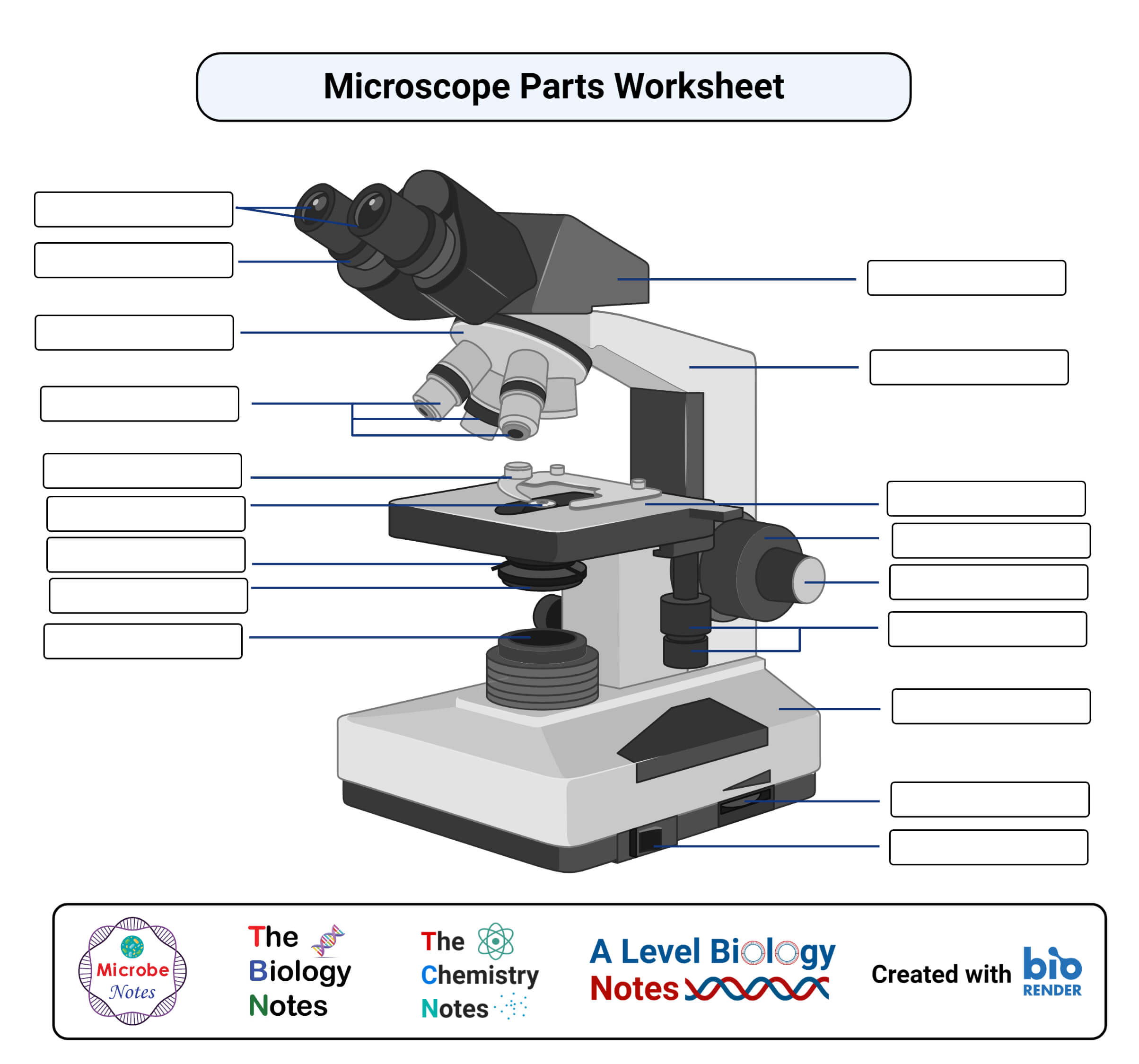
Compound Microscope Parts, Functions, and Labeled Diagram Compound Microscope Definitions for Labels Eyepiece (ocular lens) with or without Pointer: The part that is looked through at the top of the compound microscope. Eyepieces typically have a magnification between 5x & 30x. Monocular or Binocular Head: Structural support that holds & connects the eyepieces to the objective lenses.
Compound Microscope Stock Illustrations – 734 Compound ...
Microscope Parts and Functions It also allows the specimen to be labeled, transported, and stored without damage. Stage: The flat platform where the slide is placed. Stage clips: Metal clips that hold the slide in place. Stage height adjustment (Stage Control): These knobs move the stage left and right or up and down.
compound microscope with label and functions - Clip Art Library
(PDF) general-chemistry.pdf | Sumit Banerjee - Academia.edu general-chemistry.pdf
Compound Microscope Review - ppt download
Inorganic Chemistry 4th edition, Catherine Housecroft Purple acid phosphatases (PAPs) are a group of metallohydrolases that contain a dinuclear Fe(III)M(II) center (M(II) = Fe, Mn, Zn) in the active site and are able to catalyze the hydrolysis of a variety of phosphoric acid esters.

Cell Drawing Microscope - Binocular Compound Microscope ...
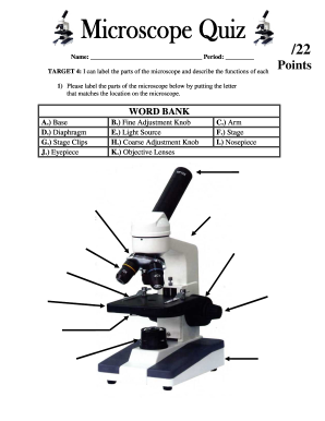
PDF Parts of a Microscope Printables - Homeschool Creations typical student microscope -other microscopes will vary) •Which part of the microscope rotates so another person can look through the eyepiece without needing to move the microscope ? the head •What is the magnification level on the eyepiece of a microscope?10x (see objective lens magnification to see how these work together)

Label Microscope Diagram - EnchantedLearning.com
Compound Microscope Parts Head/Body houses the optical parts in the upper part of the microscope. Base of the microscope supports the microscope and houses the illuminator. Arm connects to the base and supports the microscope head. It is also used to carry the microscope. When carrying a compound microscope always take care to lift it by both the arm and base ...

Microscope World Blog: What is a Compound Microscope?
Lower Secondary Science LEARNER’S BOOK 8 - Issuu Feb 22, 2021 · The photograph in Figure 1.2.1 shows a tiny part of the lungs, seen through a powerful microscope. You can see the lungs are mostly holes. These holes are called air sacs.

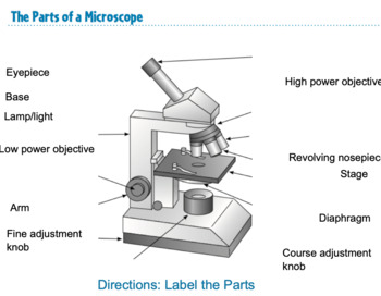
The Microscope
Compound Microscope: Definition, Diagram, Parts, Uses, Working ... - BYJUS The parts of a compound microscope can be classified into two: Non-optical parts Optical parts Non-optical parts Base The base is also known as the foot which is either U or horseshoe-shaped. It is a metallic structure that supports the entire microscope. Pillar The connection between the base and the arm are possible through the pillar. Arm

Compound Microscope Parts, Functions, and Labeled Diagram ...
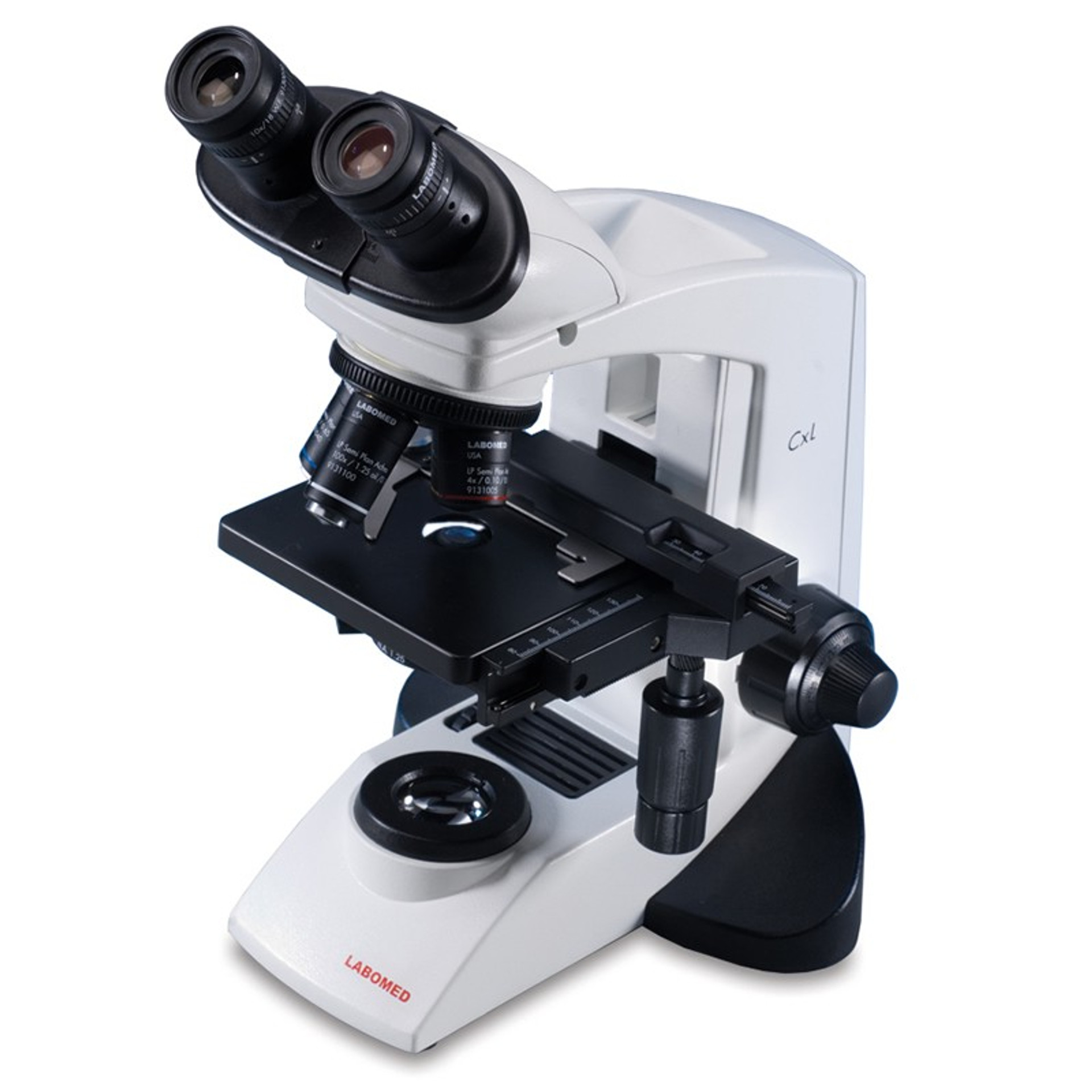
Compound Light Microscope: Everything You Need to Know A compound light microscope is a type of light microscope that uses a compound lens system, meaning, it operates through two sets of lenses to magnify the image of a specimen. It's an upright microscope that produces a two-dimensional image and has a higher magnification than a stereoscopic microscope.
Simple Microscope - Parts, Functions, Diagram and Labelling ...
Parts of a Compound Microscope and Their Functions - NotesHippo Compound microscope mechanical parts (Microscope Diagram: 2) include base or foot, pillar, arm, inclination joint, stage, clips, diaphragm, body tube, nose piece, coarse adjustment knob and fine adjustment knob. Base: It's the horseshoe-shaped base structure of microscope. All of the other components of the compound microscope are supported by it.

Label the microscope — Science Learning Hub
Compound Microscope Parts - Labeled Diagram and their Functions Labeled diagram of a compound microscope Major structural parts of a compound microscope There are three major structural parts of a compound microscope. The head includes the upper part of the microscope, which houses the most critical optical components, and the eyepiece tube of the microscope.
Parts of a Microscope with Their Functions • Microbe Online
Solved Label the image of a compound light microscope using - Chegg Expert Answer. 100% (17 ratings) Transcribed image text: Label the image of a compound light microscope using the terms provided.
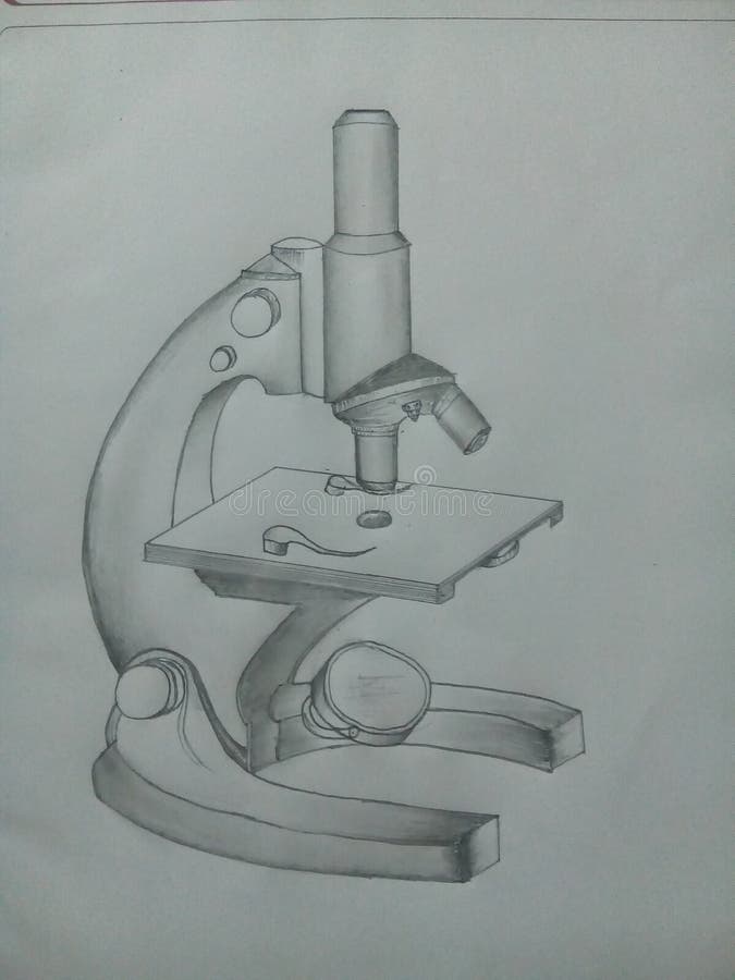
Free Microscope Drawing, Download Free Microscope Drawing png ...
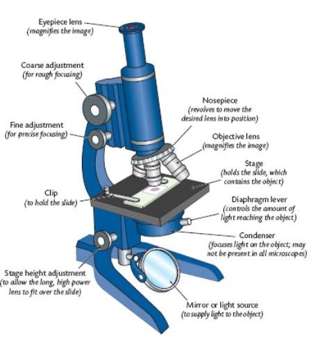
Label the microscope — Science Learning Hub Use this interactive to identify and label the main parts of a microscope. Drag and drop the text labels onto the microscope diagram. eye piece lens diaphragm or iris coarse focus adjustment stage base fine focus adjustment light source high-power objective Download Exercise Tweet
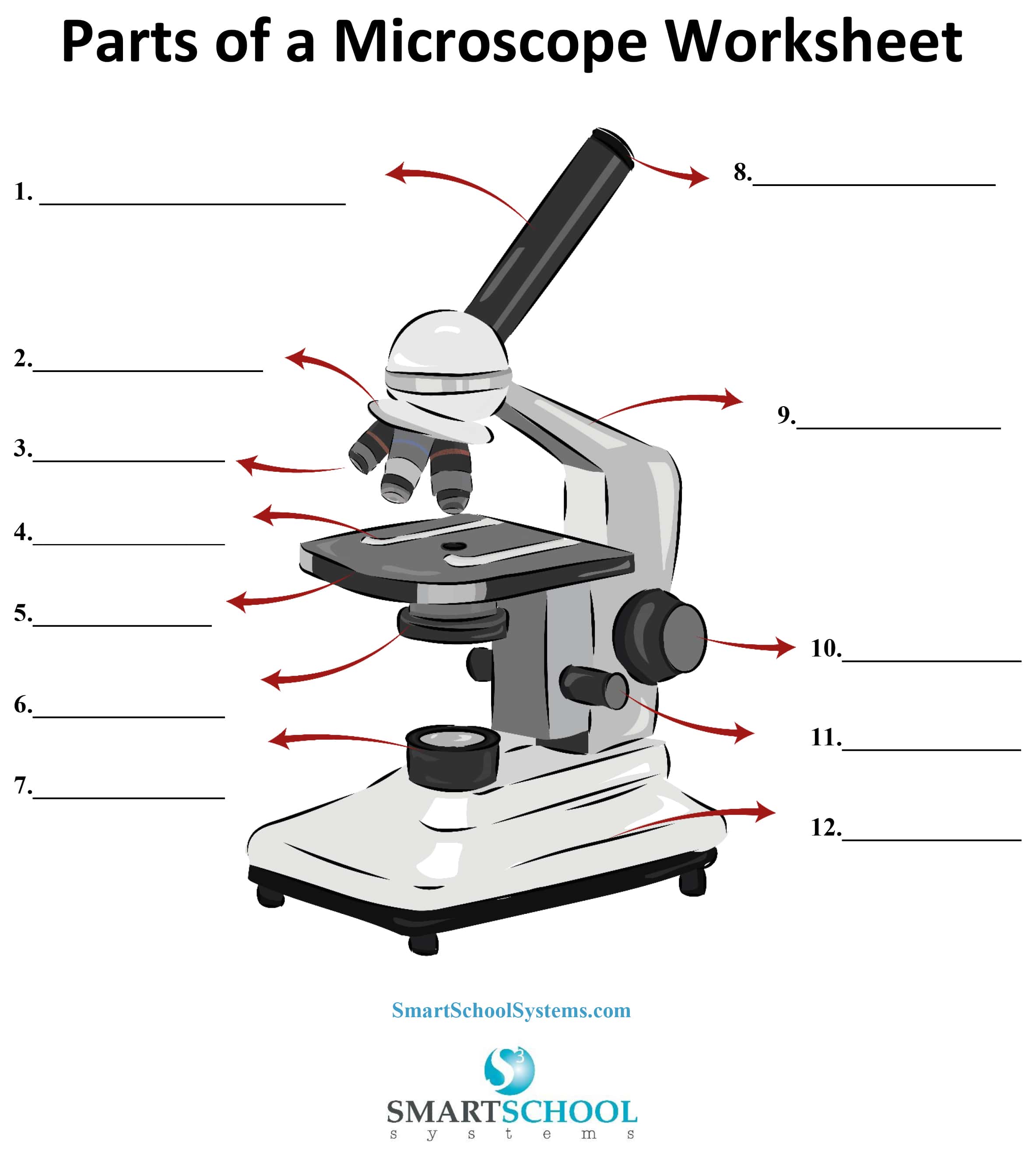
Parts of a Microscope - SmartSchool Systems
Parts of a microscope with functions and labeled diagram
Compound Light Microscope Labeled - ClipArt Best

Compound Microscope Parts – Labeled Diagram and their ...
Parts Of A Microscope Labeling Teaching Resources | TpT
How to see a plant cell under a compound microscope - Quora

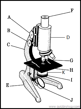
This is a common compound microscope. Label its parts from A ...

Ms. Chea's Science Class: Microscopes

microscope | Types, Parts, History, Diagram, & Facts | Britannica

Microscope Components - Science Quiz
Parts of a microscope with functions and labeled diagram
Free Microscope Drawing, Download Free Microscope Drawing png ...

Cytology. Cytology. radiation used to illuminate the specimen ...

Compound Microscope Parts

Biology 30 Compound Microscope Diagram | Quizlet

Diagram of a Compound Microscope

Label the Microscope Diagram | Download Scientific Diagram
Microscope Fill In The Blank - Fill Online, Printable ...
Post a Comment for "39 compound microscope diagram without labels"