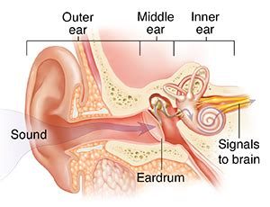
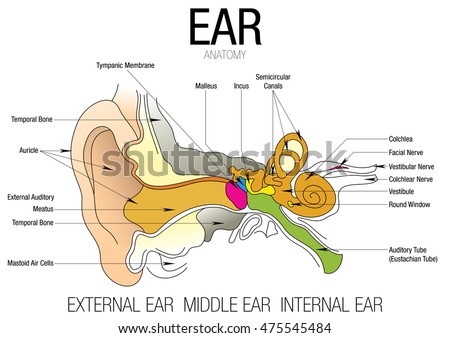
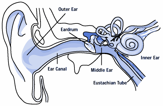
40 ear anatomy without labels
Anatomical Line Drawings - Today on Medscape Heart - sectioned view. go to drawing with labels go to drawing without labels. Heart and Lungs - anterior view. go to drawing with labels go to drawing without labels. Heart Valves - superior ... The Ear: Anatomy, Function, and Treatment - Verywell Health Essential organs of human hearing and balance, the ears are located on either side of the head, at the level of the nose. Separated into an inner, middle, and outer ear, each ear is an intricate and complicated mixture of bones, nerves, and muscles.
Parts and Components of Human Ear and Their Functions There're several parts and components of ear, which are divided into the outer, middle and inner ear sections. Each part is essential to the overall function of it. The ear parts allow the body to capture sound waves out of the air, translate them into vibrations and send these signals to the brain to be interpreted.
Ear anatomy without labels
A Guide to Different Ear Piercing Types and Their Positions The conch is the large expanse of cartilage that forms the back of the ear. A conch piercing is located in the big area of cartilage just above the earlobes and the anti-tragus on the inner ear. A ring or a barbell can be worn on this type of piercing. If selecting a ring, make sure it is large enough to encircle the outer ear. Circulatory System Diagram - New Health Advisor Coronary circuit mainly consists of cardiac veins including anterior cardiac vein, small vein, middle vein and great (large) cardiac vein. There are different types of circulatory system diagrams; some have labels while others don't. The color blue stands for deoxygenated blood while red stands for blood which is oxygenated. Working with sensor locations — MNE 1.1.dev0 documentation About montages and layouts#. Montages contain sensor positions in 3D (x, y, z in meters), which can be assigned to existing EEG/MEG data. By specifying the locations of sensors relative to the brain, Montages play an important role in computing the forward solution and inverse estimates. In contrast, Layouts are idealized 2D representations of sensor positions.
Ear anatomy without labels. Blood vessels: Histology and clinical aspects - Kenhub In the bone marrow, liver and spleen, the capillaries either have incompletely formed (or completely absent) basement membranes underlying widely spaced endothelial cells. There are usually no gap junctions between these cells and the vessel allows for direct transportation from the vascular lumen to the surrounding cells. › Atlas-Human-Anatomy-InteractiveAtlas of Human Anatomy: Including Student Consult Interactive ... Explore additional unique perspectives of difficult-to-visualize anatomy through all-new paintings by Dr. Carlos Machado, including breast lymph drainage; the pterygopalantine fossa; the middle ear; the path of the internal carotid artery; and the posterior knee, plus additional new plates on arteries of the limbs and new radiologic images. Ancient genomes from the last three millennia support multiple human ... The test groups are shown on the x and y axis labels. Data are presented as exact F 4-values ± 2 s.e. indicated by the grey lines. Linear regression lines for the individuals from the North ... Positions and Functions of the Four Brain Lobes - MD-Health.com The occipital lobe, the smallest of the four lobes of the brain, is located near the posterior region of the cerebral cortex, near the back of the skull. The occipital lobe is the primary visual processing center of the brain. Here are some other functions of the occipital lobe: Visual-spatial processing. Movement and color recognition.
Sheep - Wikipedia Sheep or domestic sheep (Ovis aries) are domesticated, ruminant mammals typically kept as livestock.Although the term sheep can apply to other species in the genus Ovis, in everyday usage it almost always refers to Ovis aries.Like all ruminants, sheep are members of the order Artiodactyla, the even-toed ungulates.Numbering a little over one billion, domestic sheep are also the most numerous ... Female reproductive organs: Anatomy and functions | Kenhub The uterus ( womb) is a hollow muscular organ located deep within the pelvic cavity. Anterior to the rectum and posterosuperiorly to the urinary bladder, the uterus normally sits in a position of anteversion and anteflexion. The endometrial lining of the uterus proliferates each month in preparation for embryo implantation. All Otitis Is Not Created Equal: AOM vs OME - Medscape Otitis media with effusion (OME) is fluid in the middle ear without signs or symptoms of inflammation that can occur just prior to or persist after an infection for a few days or up to many weeks. OME is very common in young children. About 90% of children have OME at some time before school age, most often between ages 6 months and 4 years. [3] › cefdinir › articleCefdinir Antibiotic Side Effects, Uses (Strep, Middle Ear ... Cefdinir is an antibiotic in the cephalosporin drug class prescribed to treat infections, for example, middle ear, tonsillitis, strep throat, bronchitis, and sinusitis. Common side effects are nausea, abdominal pain, loose stools, and vaginitis. Dosage and pregnancy and breastfeeding safety information are included.
Anatomical Line Drawings - Today on Medscape Male Reproductive Organs - sagittal section. go to drawing with labels go to drawing without labels. Male Reproductive Organs - cross section. go to drawing with labels go to drawing without ... Examining the Ears, Nose, and Oral Cavity in the Older Patient Place the vibrating tuning fork on the patient's forehead at an equal distance between the ears and ask whether they hear the sound (not feel the vibration). If they cannot hear it at all, then they have bilateral sensorineural hearing loss and, in fact, probably cannot hear your voice. Review: LACMA strikes gold with Indigenous Colombia exhibit - Los ... In the center of the chest is a heart-shaped breastplate trimmed in geometric shapes, in the middle of which is a chunky mask sporting its own extravagant nose and ear ornaments. Finally, simple... Neck Space Anatomy | SpringerLink Head and neck anatomy is conventionally divided into the naso-, oro-, and hypopharynx (Fig. 1), along with the oral cavity.While this approach has its merits, particularly with regard to preferred patterns of metastatic nodal spread from primary head and neck squamous cell carcinoma (SCC), it erroneously suggests that they are discrete areas when if fact they form one continuous naso-digestive ...
Ramsey syndrome - Mohammed Woodson locked-in syndrome lis also known as pseudocoma is a condition in which a patient is aware but cannot move or communicate verbally due to complete paralysis of nearly all voluntary muscles in the body except for vertical eye movements and blinkingthe individual is conscious and sufficiently intact cognitively to be able to communicate with eye …
› 40600784 › LIBRO_PARA_COLOREAR_NETTER(PDF) LIBRO PARA COLOREAR NETTER - Academia.edu Enter the email address you signed up with and we'll email you a reset link.
An Introduction to the 5 Layers of Abdominal Wall - New Health Advisor From deep to superficial, the anatomic layers that create the layers of abdominal wall are: Peritoneum. Extraperitoneal fascia (deep fascia) Muscle. Subcutaneous tissue (superficial fascia) Skin. It will depend on the location whether different layers are absent or present. 1.
LACMA STRIKES GOLD - PressReader It's flanked by a matching pair of huge, dish-shaped ear ornaments. In the center of the chest is a heartshaped breastplate trimmed in geometric shapes, in the middle of which is a chunky mask sporting its own extravagant nose and ear ornaments. Finally, simple gold cuffs wrap the wrists and ankles, while a ring encircles a finger.
Arthropod - Wikipedia Arthropods form the phylum Arthropoda. They are distinguished by their jointed limbs and cuticle made of chitin, often mineralised with calcium carbonate. The arthropod body plan consists of segments, each with a pair of appendages. Arthropods are bilaterally symmetrical and their body possesses an external skeleton.
› Flents-Quiet-Contour-Plugs-PairFlents Ear Plugs, 10 Pair with Case, Ear Plugs for Sleeping ... Flents Ear Plugs, 55 Pair, Ear Plugs for Sleeping, Snoring, Loud Noise, Traveling, Concerts, Construction, & Studying, Contour to Ear, NRR 33, Made in the USA targeal Earplugs,Noise Cancelling Earplugs with Portable PVC Case, Highest NRR 33dB Soft Form Earplugs, Reusable Sound Blocking Earplugs for Snoring, Travel, Work, Studying-4
› enAnatomy, medical imaging and e-learning for ... - IMAIOS IMAIOS and selected third parties, use cookies or similar technologies, in particular for audience measurement. Cookies allow us to analyze and store information such as the characteristics of your device as well as certain personal data (e.g., IP addresses, navigation, usage or geolocation data, unique identifiers).
OSH Answers - Canadian Centre for Occupational Health and Safety Follow manufacturer's instructions. With earplugs, for example, the ear should be pulled outward and upward with the opposite hand to enlarge and straighten the ear canal, and insert the plug with clean hands. Ensure the hearing protector tightly seals within the ear canal or against the side of the head. Hair and clothing should not be in the way.
Skull Head Anatomy Sticker | Etsy This skull was created due to my love for anatomy and neurology. It was made for anyone who appreciates this complex science, even if you are in the healthcare field or not! Any item can be exciting with this fun sticker! Add a little extra motivation and joy to your life with these durable vinyl
Room - Chapter 5 - teammaddison - Grey's Anatomy [Archive of Our Own] Get back in Wardrobe, go to sleep.". I beg, I try to go to him, but Derek pushes me away. I fall hard to the ground, hitting my head on the bedside table. I move my hand to my head, it's bleeding. I try to get up, to go to Emmerson again, but Derek looks at me dangerously. "Stay down.". He warns.
› en › e-AnatomyPetrous bone CT: normal anatomy| e-Anatomy - IMAIOS Sep 13, 2021 · Anatomy of the temporal bone: how to view the anatomical labels. This module is a comprehensive and affordable learning tool for residents and medical students and specially for neuroradiologists and otolaryngologists. It provides images in the axial and coronal planes, allowing the user to review and learn anatomy interactively.
Episodes, Showtimes, Video and more - Daily Mail TV DailyMailTV bring the best of DailyMail.com to life on television, with an edgy, fast-paced daily show featuring the hottest news headlines, celebrity breaking news and trending topic from around ...
Salvador: Dissecting the Anatomy of Someone I Both Love and Hate You stood up and looked at me, not adoringly as you did at night, not detached as you were in the mornings, but somehow repulsed and terrified. You stood up and left the room. I sat down and cried ...
Ramsay Hunt syndrome - arronkenzee.blogspot.com Ramsay Hunt syndrome type 1 also called Ramsay Hunt cerebellar syndrome is a rare form of cerebellar degeneration which involves myoclonic epilepsy progressive ataxia tremor and a dementing process. Antiviral medicines can also be given. Ramsay Hunt syndrome can be treated with anti-inflammatory drugs such as steroids.
› en › libraryAnatomy coloring books: How to use & free PDF - Kenhub Sep 30, 2021 · Generally, an anatomy coloring book will divide subject matter into sections, with each section containing many topics. For each topic you will find black and white anatomical drawings, often accompanied by labels, related text and terminology. Tired of keeping track of so many study materials?
Ramsay Hunt syndrome - Voncile Washburn Ramsay Hunt syndrome type 1 also called Ramsay Hunt cerebellar syndrome is a rare form of cerebellar degeneration which involves myoclonic epilepsy progressive ataxia tremor and a dementing process. It is sometimes called herpes zoster oticus. Shingles affecting either ear is a condition caused by a virus called herpes zoster oticus.
KevinMD.com | Social media's leading physician voice Physician. June 12, 2022. In the mid 16th century, surgical education was that of a true apprenticeship. The student learned through direct observership and imitation of a skilled, elder surgeon. It wasn't until the beginning of the 20th century that surgical education evolved into a formalized and structured program. Dr.


Post a Comment for "40 ear anatomy without labels"