38 diagram of the human eye without labels
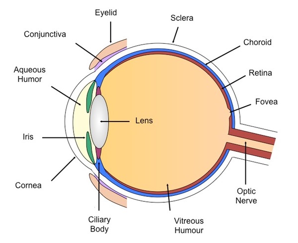
Human Body Diagram - Bodytomy The head, neck, torso, a pair of arms and legs, respectively constitute the external view of the body, often described as the superficial, first-layer of the human body. However, internally, the structure is far complex and intricate. Know that there are 11 organ systems of the body: Circulatory System, Respiratory System, Immune System ... Eye Diagram With Labels and detailed description - BYJUS A brief description of the eye along with a well-labelled diagram is given below for reference. Well-Labelled Diagram of Eye The anterior chamber of the eye is the space between the cornea and the iris and is filled with a lubricating fluid, aqueous humour. The vascular layer of the eye, known as the choroid contains the connective tissue.
Draw a labeled diagram of human eye. Write the functions of Cornea ... Write the functions of Cornea, Iris, Pupil, eye lens and retina. - 239561 p4unshaAishivio p4unshaAishivio 10.12.2015 Science Secondary School answered • expert verified Draw a labeled diagram of human eye. Write the functions of Cornea, Iris, Pupil, eye lens and retina. ... This is to ensure there is enough light for vision without damaging ...

Diagram of the human eye without labels
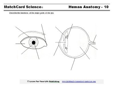
Eye anatomy: A closer look at the parts of the eye In a number of ways, the human eye works much like a digital camera: Light is focused primarily by the cornea — the clear front surface of the eye, which acts like a camera lens. The iris of the eye functions like the diaphragm of a camera, controlling the amount of light reaching the back of the eye by automatically adjusting the size of the ... Anatomy of the eye: Quizzes and diagrams - Kenhub Oct 28, 2021 · Take a look at the diagram of the eyeball above. Here you can see all of the main structures in this area. Spend some time reviewing the name and location of each one, then try to label the eye yourself - without peeking! - using the eye diagram (blank) below. Unlabeled diagram of the eye Label the Eye Worksheet - Teacher-Made Learning Resources - Twinkl The first page is a labelling exercise with two diagrams of the human eye. One is a view from the outside, and the other is a more detailed cross-section. On the second page, you'll find a set of answers showing the properly labelled human eyes, designed to help you check the worksheets without having to come up with your own answer key.
Diagram of the human eye without labels. Label the Eye Diagram - Enchanted Learning Label the Eye Diagram. Human Anatomy. Read the definitions, then label the eye anatomy diagram below. Cornea - the clear, dome-shaped tissue covering the front of the eye. Iris - the colored part of the eye - it controls the amount of light that enters the eye by changing the size of the pupil. Lens - a crystalline structure located just behind ... Eye Diagram Unlabelled - schematron.org Best Human eye diagram unlabelled free vector download for commercial use in ai, eps, cdr, svg vector illustration graphic art design format. human eye. Ask A Biologistcoloring page | Web address:schematron.org coloring. Human Eye. Page 2. 5. 3. 2. 4. How to draw human eye in easy steps -10th -Physics - science - CBSE syllabus - NCERT class 10 File:Diagram of human eye without labels.svg - Wikimedia Commons File:Diagram of human eye without labels.svg. Size of this PNG preview of this SVG file: 410 × 430 pixels. Other resolutions: 229 × 240 pixels | 458 × 480 pixels | 732 × 768 pixels | 976 × 1,024 pixels | 1,953 × 2,048 pixels. Human Ear Diagram - Bodytomy The Structure of Human Ear. Helix: It is the prominent outer rim of the external ear. Antihelix: It is the cartilage curve that is situated parallel to the helix. Crus of the Helix: It is the landmark of the outer ear, situated right above the pointy protrusion known as the tragus. Auditory Ossicles: The three small bones in the middle ear ...
Activity Sheet 1: How the Eyes Work | Human eye diagram, Teaching ... Description Use these simple eye diagrams to help students learn about the human eye. Three differentiated worksheets are included: 1. Write the words using a word bank 2. Cut and paste the words 3. Write the words without a word bank Labels include: eyebrow, eyelid, eyelashes, pupil, iris, and sclera. Eye Anatomy: Parts of the Human Eye - Vision Center Apr 05, 2022 · The lens of the eye (or crystalline lens) is the transparent lentil-shaped structure inside your eye. This is the natural lens. It is located behind the iris and to the front of the vitreous humor (vitreous body). The vitreous humor is a clear, colorless, gelatinous mass that fills the gap between the lens and the retina in the eye. Label Parts of the Human Eye - University of Dayton Label Parts of the Human Eye. Select One Anterior Chamber Ciliary Body Cornea Fibrous Tunic Iris Lateral Rectus Muscle Lens Medial Rectus Muscle Optic Disk Optic Nerve Pupil Retina Vascular Tunic Vitreous Nerve. Select One Anterior Chamber Ciliary Body Cornea Fibrous Tunic Iris Lateral Rectus Muscle Lens Medial Rectus Muscle Optic Disk Optic ... The Eyes (Human Anatomy): Diagram, Optic Nerve, Iris, Cornea ... - WebMD Your eye is a slightly asymmetrical globe, about an inch in diameter. The front part (what you see in the mirror) includes: Iris: the colored part. Cornea: a clear dome over the iris. Pupil: the ...
Diagram of the Eye - Home - Lions Eye Institute To understand the eye and its functions, it’s important to understand how the eye works, see below diagrams for both the external eye and the internal eye. The External Eye Instructions Click the parts of the eye to see a description for each. Hover the diagram to zoom. The Internal Eye Instructions File:Schematic diagram of the human eye en.svg - Wikimedia Diagram of the human eye in English. It shows the lower part of the right eye after a central and horizontal section. ... Full redraw: Group labels in accordance with the "Foundational Model Explorer." Added "Macula" and "Uvea" and removed "Zonular fibres." ... File:Diagram of human eye without labels.svg; File:Figure of diplopia perception ... Blank Eye Diagram - Healthiack Blank Eye Diagram This short article displays Blank Eye Diagram. Please click on the image (s) to view larger version. Feel free to browse our website for more information on this particular topic. Best viewed on 1280 x 768 px resolution in any modern browser. Blank eye diagram 1063 Blank eye diagram 1020 Blank eye diagram 1023 Eye Diagram - Differentiated Worksheets and EASEL Activities - Pinterest Eye Diagram - Differentiated Worksheets and EASEL Activities Description Use these simple eye diagrams to help students learn about the human eye. Three differentiated worksheets are included: 1. Write the words using a word bank 2. Cut and paste the words 3.
Food related medical terms: July 2011
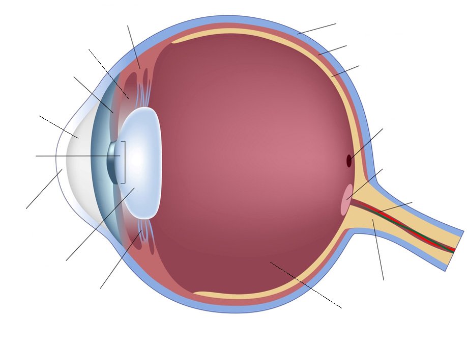
Cross sectional diagram of human eye [1]. | Download Scientific Diagram A total of 100 photos were included in the analysis—50 sick and 50 normal eyes. Small lesions in diabetic retinopathy could be automatically diagnosed by the system with an accuracy of 98%.
Human Eye Diagram
PDF Parts of the Eye - National Eye Institute | National Eye Institute Eye Diagram Handout Author: National Eye Health Education Program of the National Eye Institute, National Institutes of Health Subject: Handout illustrating parts of the eye Keywords: parts of the eye, eye diagram, vitreous gel, iris, cornea, pupil, lens, optic nerve, macula, retina Created Date: 12/16/2011 12:39:09 PM

Human Eye Anatomy Diagram Medical Description Stock Photo 231982309 - Shutterstock
Blank ear diagrams and quizzes: The fastest way to learn - Kenhub Take a moment to look at the ear model labeled above. This shows you all of the structures you've just learned about in the video, labeled on one diagram. Seeing them all together in this way is a great way to learn, since anatomical structures do not exist in isolation. That's why labeling the ear is an effective way to begin your revision.
Anatomy - Vision
BYJUS BYJUS
Anatomy Fill In The Blanks
Eye Anatomy: Parts of the Eye and How We See Behind the anterior chamber is the eye's iris (the colored part of the eye) and the dark hole in the middle called the pupil. Muscles in the iris dilate (widen) or constrict (narrow) the pupil to control the amount of light reaching the back of the eye. Directly behind the pupil sits the lens. The lens focuses light toward the back of the eye.

The Eye - Science Quiz
PDF Eye Anatomy Handout - National Eye Institute of light entering the eye. Lens: The lens is a clear part of the eye behind the iris that helps to focus light, or an image, on the retina. Macula: The macula is the small, sensitive area of the retina that gives central vision. It is located in the center of the retina. Optic nerve: The optic nerve is the largest sensory nerve of the eye.
picture front of the eye without labels clipart 20 free Cliparts | Download images on Clipground ...
Eye Diagram Teaching Resources | Teachers Pay Teachers Anatomy of the Eye Diagrams for Coloring/Labeling, with Reference and Summary by Homemade For Play 7 $1.95 PDF This printable contains 13 clear and simple cross sectional diagrams of the human eye.

Continuing Medical Education | Body anatomy, Human eye diagram, Human eye
Label Parts of the Human Ear - University of Dayton Label Parts of the Human Ear. Select One Auditory Canal Cochlea Cochlear Nerve Eustachian Tube Incus Malleus Oval Window Pinna Round Window Semicircular Canals Stapes Tympanic Membrane Vestibular Nerve. Select One Auditory Canal Cochlea Cochlear Nerve Eustachian Tube Incus Malleus Oval Window Pinna Round Window Semicircular Canals Stapes ...

File:Diagram of human eye without labels.svg - Wikimedia Commons
File:Schematic diagram of the human eye no.svg - Wikipedia Jul 13, 2021 · Original upload log []. This image is a derivative work of the following images: File:Schematic diagram of the human eye en.svg licensed with PD-self 2008-02-02T01:33:45Z Jakov 508x516 (54267 Bytes) suspensory ligament, arrow was wrong

Eye Diagram Without Labels | via Anatomy Pictures Gallery if… | Flickr
Label the Eye Worksheet - Teacher-Made Learning Resources - Twinkl The first page is a labelling exercise with two diagrams of the human eye. One is a view from the outside, and the other is a more detailed cross-section. On the second page, you'll find a set of answers showing the properly labelled human eyes, designed to help you check the worksheets without having to come up with your own answer key.

Notes of Ch 11 Human Eye and Colourful World| Class 10th Science « Study Rankers
Anatomy of the eye: Quizzes and diagrams - Kenhub Oct 28, 2021 · Take a look at the diagram of the eyeball above. Here you can see all of the main structures in this area. Spend some time reviewing the name and location of each one, then try to label the eye yourself - without peeking! - using the eye diagram (blank) below. Unlabeled diagram of the eye

28 best Human Body 5th Grade images on Pinterest | School, Teaching science and The brain
Eye anatomy: A closer look at the parts of the eye In a number of ways, the human eye works much like a digital camera: Light is focused primarily by the cornea — the clear front surface of the eye, which acts like a camera lens. The iris of the eye functions like the diaphragm of a camera, controlling the amount of light reaching the back of the eye by automatically adjusting the size of the ...

File:Schematic diagram of the human eye-es.svg - Wikimedia Commons
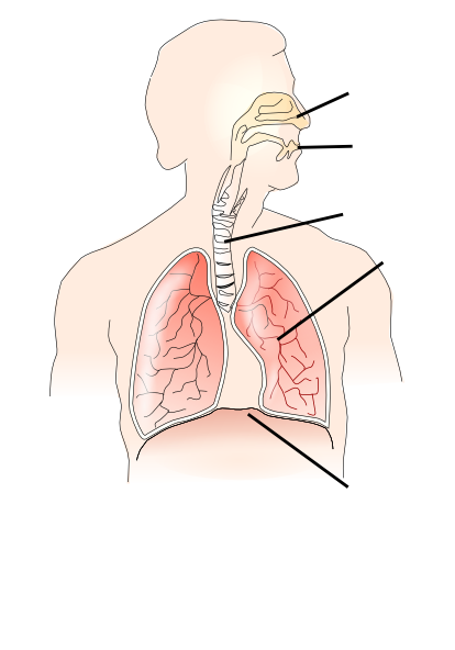
Unlabelled Respiratory System Clip Art at Clker.com - vector clip art online, royalty free ...

Activity Sheet 1: How the Eyes Work | Human eye diagram, Teaching biology, Human body activities

Diagram Of Human Eye Without Label | MedicineBTG.com
Post a Comment for "38 diagram of the human eye without labels"